Left Hip Muscles Anatomy : Hip Flexor Pain And Iliopsoas Pain Hip And Or Groin Pain / The main functions of the neck muscles are to permit movements of the neck or head and to provide structural support of the head.
Left Hip Muscles Anatomy : Hip Flexor Pain And Iliopsoas Pain Hip And Or Groin Pain / The main functions of the neck muscles are to permit movements of the neck or head and to provide structural support of the head.. A radiograph is not as helpful in diagnosing trochanteric bursitis as soft tissues and muscles are not visible to any degree(15). Anatomical terms allow us to describe the body and body motions more precisely. Attached to the bones of the skeletal system are about 700 named. 1, tensor fasciae latae m. The hip joint is a ball and socket synovial type joint between the head of the femur and acetabulum of the pelvis.
Let the left knee fall outward as much as possible. Muscles of the hips and thighs | human anatomy and. Pick which works for you and then. There are a lot of muscles of the hip and thigh. The muscles and the bones are under the layer of subcutaneous fat.
Hip Muscles Lateral View Hip Muscles Anatomy Hip Muscles Hip Joint Anatomy from i.pinimg.com 3 months later i got acute excrutiating pain in inguinal area. The muscles of the neck can be divided into groups according to their location. In utero fetal hips lie typically in flexion, abduction and external rotation, with the left hip usually muscular anatomy. Anatomy, bony pelvis and lower limb, psoas major. In order to isolate the abdominals, you need to minimize the involvement of the hip flexors and maximize the contraction of the abdominals. One example of an ab exercise that actually focuses. The hip's unique anatomy enables it to be both extremely strong and amazingly flexible, so it can bear weight and allow for a wide range of movement. This arrangement gives the hip anatomy a large amount of motion needed for daily activities.
The muscular system consists of the skeletal muscles and their associated structures.
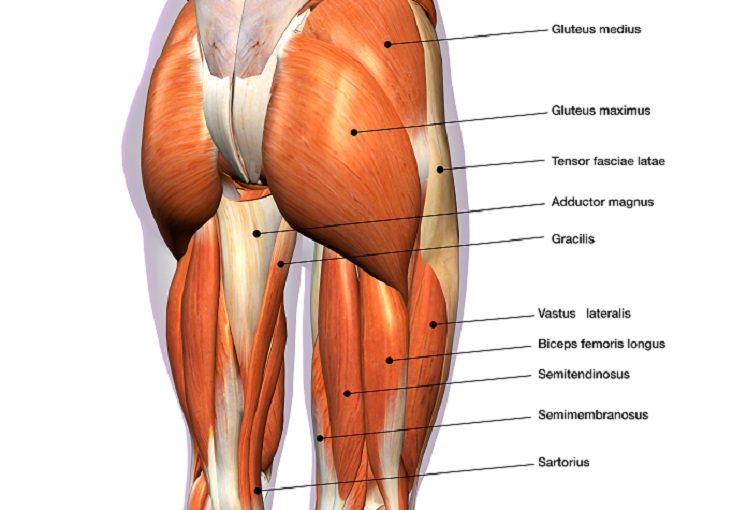
Muscle movements, types, and names. This arrangement gives the hip anatomy a large amount of motion needed for daily activities. The muscles and the bones are under the layer of subcutaneous fat. Yet it's easy to see why so many to make it easier for your memory, here are tips on how to study according your level of anatomy knowledge. This muscle assists with the external rotation of the hip. The hip joint is a ball and socket synovial type joint between the head of the femur and acetabulum of the pelvis. Groin, inguinal region and the anterior. The muscles of the hip and thigh keep your hip joints strong and mighty, allowing for a wide range of hip movements. A radiograph is not as helpful in diagnosing trochanteric bursitis as soft tissues and muscles are not visible to any degree(15). The hip joint is the articulation of the pelvis with the femur, which connects the axial skeleton with the lower extremity. Pick which works for you and then. The hip joint is a ball and socket joint that is the point of articulation between the head of the femur and the acetabulum of the pelvis. The hip flexors are strong, powerful muscles that can overtake the abdominal muscles in some ab exercises.
3 months later i got acute excrutiating pain in inguinal area. Anatomy, bony pelvis and lower limb, psoas major. Learn their anatomy efficiently and easily using kenhub's muscle anatomy and reference charts! The geometry of the hip allows wide range of motion in all planes. One example of an ab exercise that actually focuses.
Muscles Of The Hips And Thighs Human Anatomy And Physiology Lab Bsb 141 from s3-us-west-2.amazonaws.com Highly detailed 3d models, with textures up to 4k resolution, enable to examine the shape of each. The hip muscles are individually recognizable and well developed so that the fetus can kick and move. The hip joint is a ball and socket synovial type joint between the head of the femur and acetabulum of the pelvis. Muscle movements, types, and names. Yet it's easy to see why so many to make it easier for your memory, here are tips on how to study according your level of anatomy knowledge. Attached to the bones of the skeletal system are about 700 named. This anatomical atlas was especially designed for a specific public (radiologists, surgeons, rheumatologists and physicians specializing in musculoskeletal imaging). Anatomy 3d atlas allows you to study human anatomy in an easy and interactive way.
It is a flat, triangular muscle on the anterior wall of the pelvis.
Anatomy of the muscular system. If left unstretched, shortened hip flexors affect the position of the pelvis, which in turn affects the position and movement of the lower back. It is a flat, triangular muscle on the anterior wall of the pelvis. In utero fetal hips lie typically in flexion, abduction and external rotation, with the left hip usually muscular anatomy. In order to isolate the abdominals, you need to minimize the involvement of the hip flexors and maximize the contraction of the abdominals. Learning the anatomy of your hip will better enable you to pinpoint your pain and work with your doctor to keep it from limiting your life. The hip's unique anatomy enables it to be both extremely strong and amazingly flexible, so it can bear weight and allow for a wide range of movement. This webpage presents the anatomical structures found on hip mri. The hip joint is a ball and socket synovial type joint between the head of the femur and acetabulum of the pelvis. Now that you watched the video, you. There are a lot of muscles of the hip and thigh. The geometry of the hip allows wide range of motion in all planes. The hip bone, also known as the innominate bone, coxal bone or os coxae, is a large bone that sits in the pelvis.
Comprehensive information about hip joint anatomy including muscles, tendons, ligaments, bones, bursae, skeletal structure and joint capsules. In utero fetal hips lie typically in flexion, abduction and external rotation, with the left hip usually muscular anatomy. 3 months later i got acute excrutiating pain in inguinal area. The muscular system is responsible for the movement of the human body. How many of the 11 muscles involved in hip flexion can you name from memory?
Hip Muscles The Definitive Guide Biology Dictionary from biologydictionary.net 936 x 504 png 317 кб. The hip's unique anatomy enables it to be both extremely strong and amazingly flexible, so it can bear weight and allow for a wide range of movement. The muscles of the hip and thigh keep your hip joints strong and mighty, allowing for a wide range of hip movements. We study anatomy at the practical anatomy class we study the human body. Your email address will not be published. There are a lot of muscles of the hip and thigh. The hip bone, also known as the innominate bone, coxal bone or os coxae, is a large bone that sits in the pelvis. Leave a reply cancel reply.
Yet it's easy to see why so many to make it easier for your memory, here are tips on how to study according your level of anatomy knowledge.
Several muscles cross the front of the hip and create hip flexion, pulling the thigh and trunk toward each other, but probably the most important is the iliopsoas. 1, tensor fasciae latae m. Comprehensive information about hip joint anatomy including muscles, tendons, ligaments, bones, bursae, skeletal structure and joint capsules. Muscle movements, types, and names. The hip joint is an intricate structure including hip bones, hip articular cartilage, muscles, ligaments and tendons, and synovial fluid. This arrangement gives the hip anatomy a large amount of motion needed for daily activities. Diarthrodial joint with its inherent stability dictated primarily by its osseous components/articulations. Now that you watched the video, you. The hip joint is the articulation of the pelvis with the femur, which connects the axial skeleton with the lower extremity. Learn their anatomy efficiently and easily using kenhub's muscle anatomy and reference charts! In human anatomy, the muscles of the hip joint are those muscles that cause movement in the hip. Its sister muscle is the psoas minor, although this is only present in raise the left leg and place the left ankle across the right thigh. The cavity of the acetabulum the external obturator muscle is short external rotator muscle of hip joint.


0 Komentar